Relieve pain and restore function
Dr. Farr offers a number of non-surgical and surgical techniques to relieve pain and restore function in the knee. Learn more about the anatomy of the knee and your treatment options, which include:
Knee Anatomy
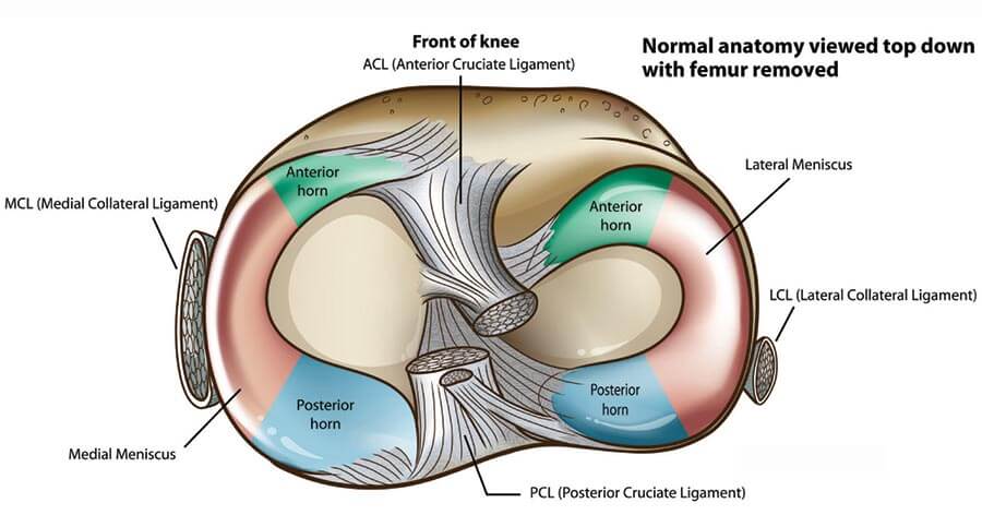
The knee joint is made up of three bones, four major ligament groups and two different types of cartilage.

Femur (Thigh Bone)
The femur portion of the knee joint has two separated parts, called condyles. Touching your knees together brings the inner condyles closer together—the medial femoral condyle (MFC). The outer condyle is the lateral femoral condyle (LFC).
Tibia (Shin Bone)
The tibia meets the femur at the knee in two areas on which the two femoral condyles "ride." These are the medial and the lateral tibial plateaus (MTP, LTP).
Patella (Knee Cap)
The patella rides in a shallow groove (or sulcus) over the front part of the femur, also called the trochlea.
Articular Cartilage
The ends of all the bones at the joint are covered by articular cartilage. This is a glistening white substance that has the consistency of firm rubber. However, it is actually a mixture of collagen and special large, sponge-like molecules that are maintained by living cells (chondrocytes). When joint fluid is normal and balanced, the surface is more slippery than water on ice. This combination allows for normal, smooth, and natural joint motion.

Meniscal Cartilage
The other type of knee cartilage is meniscus cartilage. There are two C-shaped pads which are found between the thigh and shin bones—one on each side. The medial meniscus is found on the inner knee and the lateral meniscus is on the outer aspect of the knee. The medial and lateral menisci are attached predominantly to the tibial plateaus and they serve to cushion the joint and transfer the load by assisting in distributing joint forces over a larger area. (See "Meniscus Function Analogy" below).
You may now appreciate that injury of either type of cartilage can upset the normal function of the joint. However it is important to note that this "injury" is not limited to trauma alone. It can occur with normal daily activities. Once the delicate balance of the knee is upset, the resulting abnormal loads then lead to "overload damage." Over time, initially small defects in the articular cartilage or tears in the meniscal cartilage can progress. This gradual deterioration can lead to degenerative joint disease—a form of osteoarthritis.
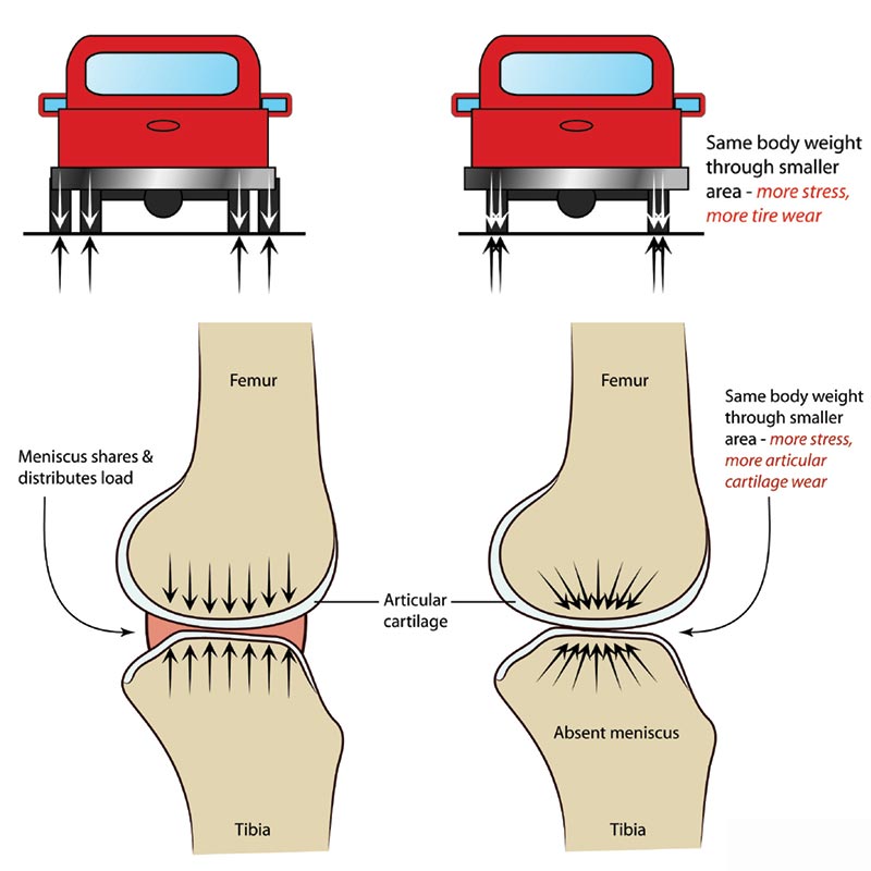
Meniscus Function Analogy
A truck with four rear tires distributes the load over a larger area than a truck carrying the same load, but with two rear tires. The two tires are stressed more than the four tires and will have greater wear. The meniscus helps distribute the body weight at the knee joint over a larger area. If the meniscus is removed, the same weight is now distributed over a smaller area and the stress increases. This increased joint stress may lead to pain and/or articular cartilage wear.